English narration · English + 中文 subtitles burned in · 英語ナレーション・英語+中文字幕 burning-in
9.1
The gas exchange system · 気体交換系
Syllabus · シラバス
English
describe the structure of the human gas exchange system, limited to: • lungs • trachea • bronchi • bronchioles • alveoli • capillary network
describe the distribution in the gas exchange system of cartilage, ciliated epithelium, goblet cells, squamous epithelium of alveoli, smooth muscle and capillaries
recognise cartilage, ciliated epithelium, goblet cells, squamous epithelium of alveoli, smooth muscle and capillaries in microscope slides, photomicrographs and electron micrographs
recognise trachea, bronchi, bronchioles and alveoli in microscope slides, photomicrographs and electron micrographs and make plan diagrams of transverse sections of the walls of the trachea and bronchus
describe the functions of ciliated epithelial cells, goblet cells and mucous glands in maintaining the health of the gas exchange system
describe the functions in the gas exchange system of cartilage, smooth muscle, elastic fibres and squamous epithelium
describe gas exchange between air in the alveoli and blood in the capillaries
Source: Cambridge International syllabus · 出典: Cambridge International シラバス
English
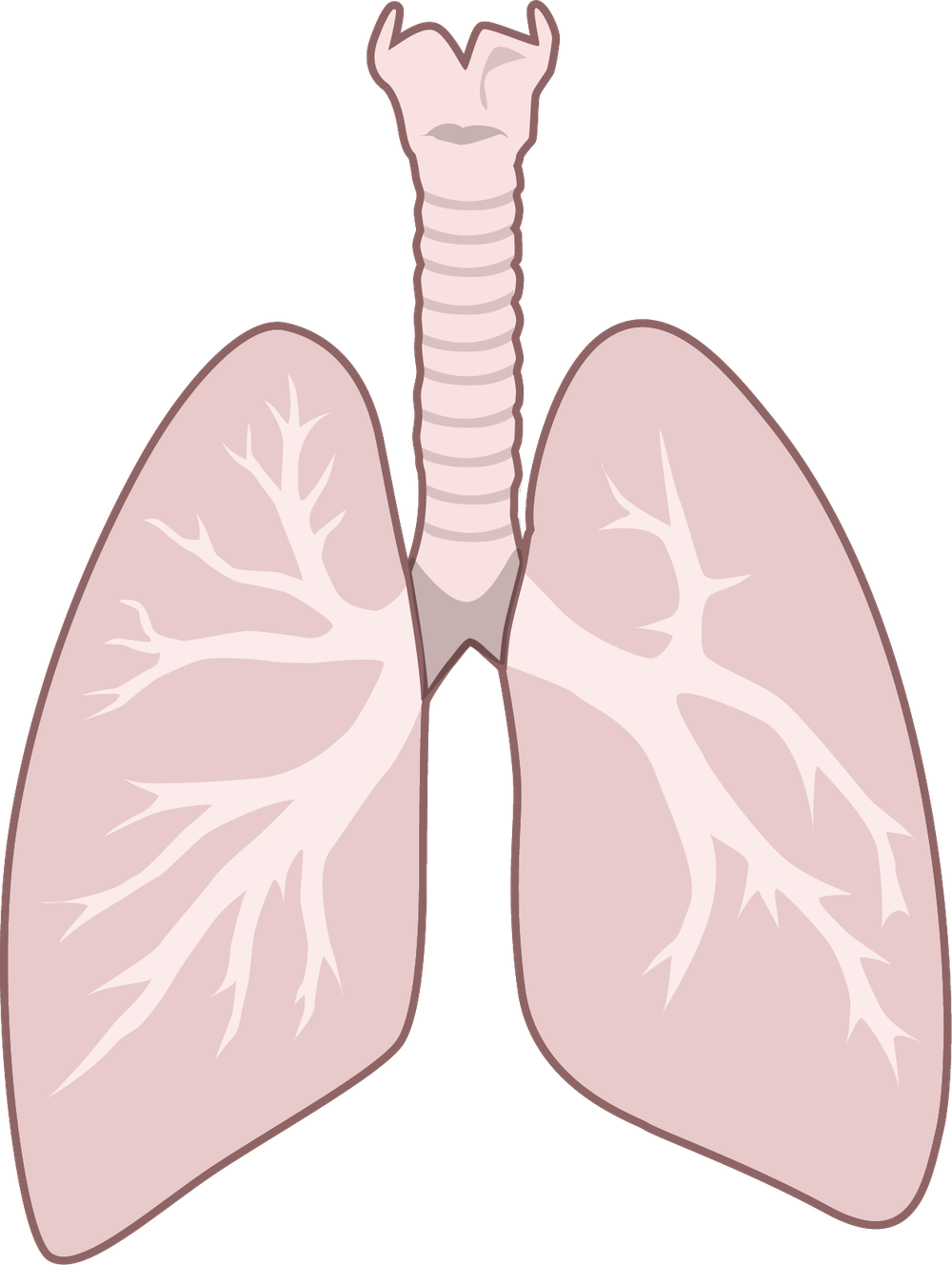
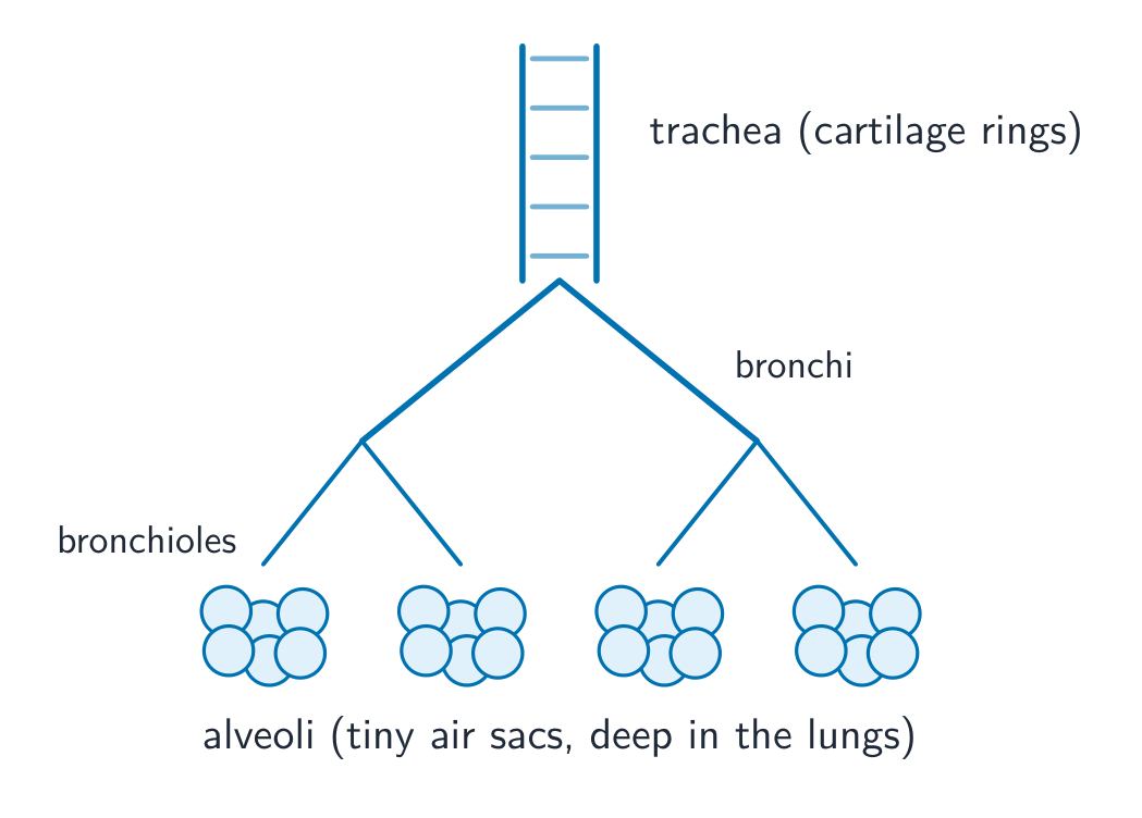
Your body needs to take in oxygen and get rid of carbon dioxide. This swap happens in the gas exchange 气体交换 system. Air follows this path into the body:
down the trachea 气管 (the windpipe),
into two bronchi 支气管 (one to each lung),
into many smaller bronchioles 细支气管,
and finally into tiny air sacs called alveoli 肺泡, deep in the lungs 肺.
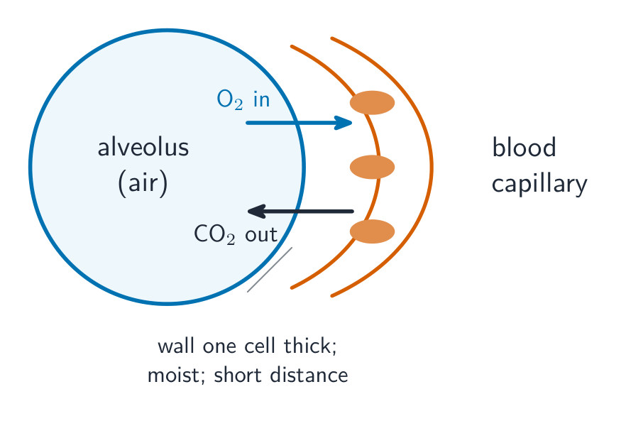
Each alveolus is wrapped in a network of capillaries 毛细血管, so air and blood are brought very close together.
Tap each part. Air branches from the trachea down to the tiny alveoli, where gas exchange happens. · 各部分をクリックしてください。空気は気管から微小な肺胞まで枝分かれし、そこでガス交換が行われます。
The tissues of the airways and what they do · 気道组织及其功能
English
Tissue
Where it is
Function
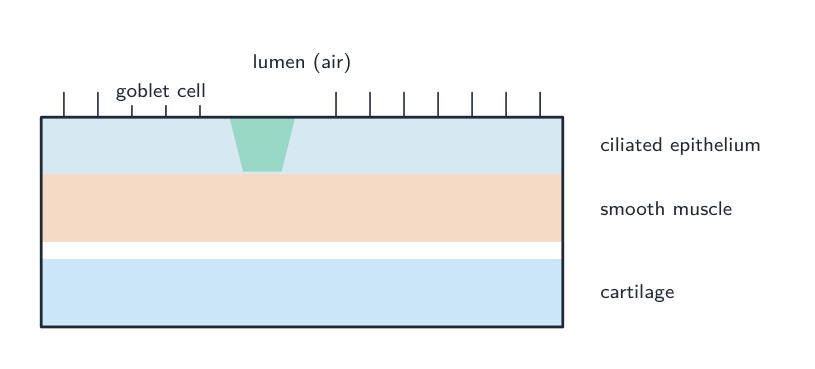
cartilage 软骨
C-shaped rings in the trachea and bronchi
holds the airway open so it cannot collapse when you breathe in
ciliated epithelium 纤毛上皮
lining the trachea and bronchi
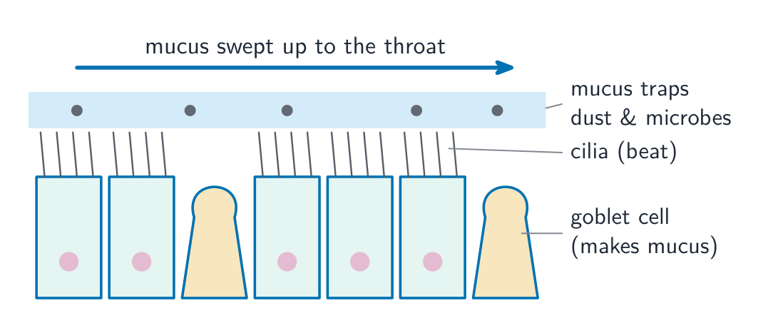
tiny hairs called cilia 纤毛 beat to sweep mucus 黏液 up towards the throat, away from the lungs
goblet cells 杯状细胞 and mucous glands 黏液腺
in the lining of the airways
make the mucus, which traps dust and microbes 微生物 that you breathe in
smooth muscle 平滑肌
in the walls of bronchi and bronchioles
contracts to make the airway narrower
elastic fibres 弹性纤维
in the airway and alveolus walls
stretch when you breathe in, then spring back to help push air out
squamous epithelium 扁平上皮
the very thin, flat lining of the alveoli
gives a very short distance for gases to cross
The cilia, goblet cells and mucous glands work together to keep the lungs clean and healthy: the mucus traps dirt and microbes, and the cilia carry it away to be swallowed.
The alveoli are excellent surfaces for exchanging gases, because they have:
a very large total surface area (millions of tiny sacs),
very thin walls — the squamous epithelium of the alveolus and the capillary wall are each only one cell thick, so the distance to cross is tiny,
a rich blood supply from the capillary network,
a moist lining, so gases dissolve before crossing.
Gases move by diffusion 扩散 down their concentration gradients 浓度梯度:
oxygen 氧气 is at a high concentration in the alveolar air and a low concentration in the blood, so it diffuses from the air into the blood.
carbon dioxide 二氧化碳 is at a high concentration in the blood and a low concentration in the alveolar air, so it diffuses from the blood into the air to be breathed out.
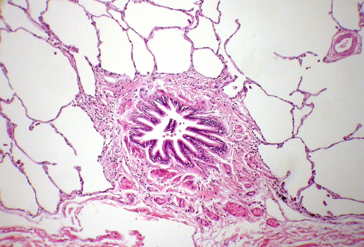
Under the microscope, real lung tissue looks like a fine pink lace. The many open spaces are the alveoli, and the thin pink lines between them are the walls where gas exchange happens:
Breathing keeps fresh air in the alveoli, and the flowing blood keeps carrying gases away. Both of these keep the concentration gradients steep, so gas exchange stays fast.
Worked example. Use Fick's law to explain why the alveoli allow such rapid gas exchange. Fick's law makes the rate of diffusion proportional to
so a fast rate needs all three terms working for it. Surface area: millions of alveoli give a huge total area, roughly $70\ \text{m}^2$. Diffusion distance: the alveolar epithelium and the capillary endothelium are each one flattened cell thick, so oxygen crosses in under $1\ \mu\text{m}$. Concentration difference: ventilation constantly refreshes the air while the circulation constantly carries oxygenated blood away, so a steep gradient is maintained. Tie each adaptation to the term of the equation it serves - listing "big surface area, thin walls, good blood supply" without linking them to Fick's law is the weaker answer.
Tap each part. Oxygen and carbon dioxide swap across a wall just one cell thick, between the air and the blood. · 各部分をクリックしてください。酸素と二酸化炭素は、空気と血液の間にある1層細胞だけの壁を介して交換されます。
Link each alveolar feature to fast diffusion: large surface area, thin (one-cell) walls, moist surface, good blood supply — a "surface area, short distance, steep gradient" answer.
Match airway tissue to function: cartilage (holds airways open), ciliated + goblet cells (trap and sweep mucus), smooth muscle and elastic fibres.
Frame answers with Fick's law ideas: rate $\propto$ surface area $\times$ concentration difference $\div$ distance.
Type to search notes, lessons, code, vocabulary and past-paper questions across every subject. · すべての科目でノートImplemented、Implemented、コード、語彙、過去問問題を検索するために入力してください。