Skip to content · ⁨Bỏ qua nội dung⁩

Transport in mammals · ⁨Vận chuyển trong động vật có vú⁩

A-Level Biology · ⁨Sinh học A-Level⁩ · Topic 8 · ⁨Chủ đề 8⁩

Video lesson for this topic · ⁨Bài học video cho chủ đề này⁩ Open the video page · ⁨Mở trang video⁩
9:17

The Circulatory System

Your heart beats about a hundred thousand times a day, and it never gets a day off. But here is the clever part. It is not one loop. It is two. Blood leaves…

English narration · English + 中文 subtitles burned in · ⁨Giọng đọc tiếng Anh · phụ đề tiếng Anh + 中文 được ghi trực tiếp⁩

8.1

The circulatory system · ⁨Hệ tuần hoàn⁩

Syllabus · ⁨Chương trình⁩
English
  1. state that the mammalian circulatory system is a closed double circulation consisting of a heart, blood and blood vessels including arteries, arterioles, capillaries, venules and veins
  2. describe the functions of the main blood vessels of the pulmonary and systemic circulations, limited to pulmonary artery, pulmonary vein, aorta and vena cava
  3. recognise arteries, veins and capillaries from microscope slides, photomicrographs and electron micrographs and make plan diagrams showing the structure of arteries and veins in transverse section (TS) and longitudinal section (LS)
  4. explain how the structure of muscular arteries, elastic arteries, veins and capillaries are each related to their functions
  5. recognise and draw red blood cells, monocytes, neutrophils and lymphocytes from microscope slides, photomicrographs and electron micrographs
  6. state that water is the main component of blood and tissue fluid and relate the properties of water to its role in transport in mammals, limited to solvent action and high specific heat capacity
  7. state the functions of tissue fluid and describe the formation of tissue fluid in a capillary network
Tiếng Việt
  1. nêu rằng hệ tuần hoàn động vật có vú là một chu kỳ kép (double circulation) kín bao gồm tim, máu và các mạch máu bao gồm động mạch (arteries), tiểu động mạch (arterioles), mao mạch (capillaries), tiểu静脉 (venules) và tĩnh mạch (veins)
  2. mô tả chức năng của các mạch máu chính trong chu kỳ phổi (pulmonary) và chu kỳ toàn thân (systemic), giới hạn ở phổi động mạch (pulmonary artery), phổi tĩnh mạch (pulmonary vein), aorta và tĩnh mạch chủ (vena cava)
  3. nhận diện động mạch, tĩnh mạch và mao mạch từ tiêu bản kính hiển vi, ảnh chụp kính hiển vi và ảnh chụp điện tử và vẽ sơ đồ cấu trúc của động mạch và tĩnh mạch trong mặt cắt ngang (TS) và mặt cắt dọc (LS)
  4. giải thích cách cấu trúc của động mạch cơ, động mạch đàn hồi, tĩnh mạch và mao mạch tương ứng liên hệ với chức năng của chúng
  5. nhận diện và vẽ tế bào hồng cầu (red blood cells), tế bào đơn nhân (monocytes), bản trung tính (neutrophils) và tế bào lympho (lymphocytes) từ tiêu bản kính hiển vi, ảnh chụp kính hiển vi và ảnh chụp điện tử
  6. nêu rằng nước là thành phần chính của máu và dịch mô và liên hệ các tính chất của nước với vai trò vận chuyển ở động vật có vú, giới hạn ở tác dụng dung môi và nhiệt dung riêng cao
  7. nêu chức năng của dịch mô (tissue fluid) và mô tả quá trình hình thành dịch mô trong mạng lưới mao mạch (capillary network)

Source: Cambridge International syllabus · ⁨Nguồn: Chương trình Cambridge International⁩

English

Mammals have a closed double circulation. "Closed" means the blood stays inside blood vessels 血管 the whole time. "Double" means the blood passes through the heart 心脏 twice for each full trip around the body. This gives two linked loops, so we call it a double circulation 双循环 and the whole thing a circulatory system 循环系统:

  • the pulmonary circulation 肺循环 carries blood from the heart to the lungs and back.
  • the systemic circulation 体循环 carries blood from the heart to the rest of the body and back.

Blood travels in this order: arteries 动脉 → arterioles 小动脉 → capillaries 毛细血管 → venules 小静脉 → veins 静脉.

The main vessels are:

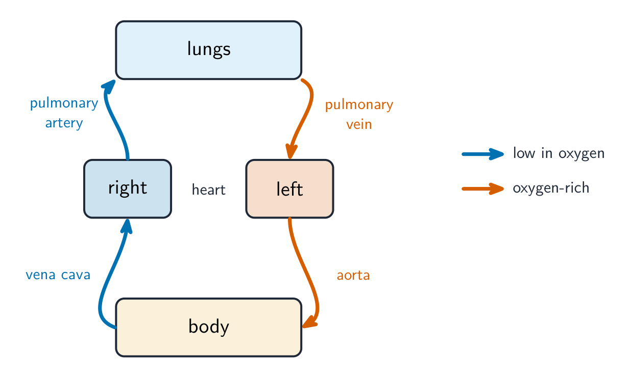
  • the pulmonary artery 肺动脉 — carries blood low in oxygen 氧气 from the heart to the lungs.
  • the pulmonary vein 肺静脉 — carries oxygen-rich blood from the lungs back to the heart.
  • the aorta 主动脉 — the big artery that carries oxygen-rich blood from the heart to the body.
  • the vena cava 腔静脉 — the big vein that returns oxygen-poor blood from the body to the heart.

How the vessels suit their jobs

Vessel Structure Function
artery thick wall of muscle 肌肉 and elastic 弹性 fibres; narrow lumen 管腔 (the space inside) carries blood at high pressure away from the heart; elastic walls stretch and recoil to smooth the flow; muscle controls the flow
capillary wall just one cell thick; very narrow short distance for exchange of substances between blood and cells
vein thin wall; wide lumen; has valves 瓣膜 returns blood at low pressure to the heart; valves stop blood flowing backwards

Blood cells

You should recognise: red blood cells 红细胞 (which carry oxygen), and three white blood cells — monocytes 单核细胞, neutrophils 中性粒细胞 and lymphocytes 淋巴细胞.

Water, plasma and tissue fluid

Water is the main part of blood. It is a good solvent 溶剂, so it carries dissolved substances, and it has a high specific heat capacity 比热容, so the blood's temperature stays steady.

At the start of a capillary, the high blood pressure pushes liquid (but not the cells or large proteins) out of the plasma 血浆 and through the capillary wall. This liquid around the cells is tissue fluid 组织液. It supplies the cells with oxygen and glucose and carries waste away. Most of it returns to the capillary at the far end, where the pressure is lower.

Tiếng Việt

Động vật có vú có tuần hoàn kép kín. "Kín" có nghĩa là máu luôn ở bên trong mạch máu. "Kép" có nghĩa là máu đi qua tim hai lần cho mỗi vòng đi hết cơ thể. Điều này tạo thành hai vòng khép kín liên kết, vì vậy ta gọi nó là tuần hoàn kép và toàn bộ hệ thống là hệ tuần hoàn:

  • chu kỳ phổi vận chuyển máu từ tim đến phổi và quay trở lại.
  • chu kỳ hệ thống vận chuyển máu từ tim đến phần còn lại của cơ thể và quay trở lại.

Máu di chuyển theo thứ tự: động mạch → tiểu động mạch → mao mạch → tiểu tĩnh mạch → tĩnh mạch.

Sơ đồ vòng lặp tuần hoàn kép: tim phải bơm máu thiếu oxy đến phổi và quay về tim trái, tim trái bơm máu giàu oxy đến cơ thể và quay về tim phải
Tuần hoàn kép: chu kỳ phổi đi đến phổi, chu kỳ hệ thống đi đến cơ thể; máu đi qua tim hai lần

Các mạch máu chính là:

  • động mạch phổi — mang máu thiếu oxy từ tim đến phổi.
  • tĩnh mạch phổi — mang máu giàu oxy từ phổi quay về tim.
  • aorta — động mạch lớn mang máu giàu oxy từ tim đến cơ thể.
  • tĩnh mạch chủ — tĩnh mạch lớn trả máu thiếu oxy từ cơ thể về tim.

Cách các mạch phù hợp với nhiệm vụ của chúng

Mạch Cấu trúc Chức năng
động mạch thành dày gồm cơ và sợi đàn hồi; lòng hẹp (khoảng trống bên trong) mang máu dưới áp suất cao ra khỏi tim; thành đàn hồi co giãn và đàn hồi lại để làm mượt dòng chảy; cơ điều khiển dòng chảy
mao mạch thành chỉ dày một lớp tế bào; rất hẹp khoảng cách ngắn để trao đổi chất giữa máu và tế bào
tĩnh mạch thành mỏng; lòng rộng; có van trả máu dưới áp suất thấp về tim; van ngăn máu chảy ngược
Cắt ngang của một động mạch có thành dày và lòng hẹp, một tĩnh mạch có thành mỏng, lòng rộng và một van, cùng một mao mạch nhỏ chỉ dày một lớp tế bào
Một động mạch có thành dày và lòng hẹp; một tĩnh mạch có thành mỏng, lòng rộng và van; một mao mạch dày một lớp tế bào

Các tế bào máu

Bạn cần nhận biết: tế bào hồng cầu (mang oxy), và ba loại tr bạch cầu — monocyte, neutrophil và lymphocyte.

Mô Blood smear nhuộm màu nhìn thấy dưới kính hiển vi quang học: rất nhiều tế bào hồng cầu nhỏ, tròn có tâm nhạt, một lymphocyte lớn có nhân tím đậm gần trung tâm, và hai neutrophil có nhân phân thùy phía dưới
Mô blood smear nhuộm màu: nhiều tế bào hồng cầu nhỏ, cộng thêm một lymphocyte (ở trung tâm) và neutrophil (nhân phân thùy) — các tế bào trắng lớn hơn và có nhân

Nước, huyết tương và dịch mô

Nước là thành phần chính của máu. Nó là dung môi tốt, do đó mang các chất tan, và có nhiệt dung riêng cao, nên nhiệt độ của máu duy trì ổn định.

Ở đầu mao mạch, áp suất máu cao đẩy chất lỏng (nhưng không phải tế bào hay protein lớn) ra khỏi huyết tương và xuyên qua thành mao mạch. Chất lỏng bao quanh tế bào này là dịch mô. Nó cung cấp oxy và glucose cho tế bào và mang chất thải đi. Phần lớn dịch này trở lại mao mạch ở cuối xa, nơi áp suất thấp hơn.

Một mao mạch với chất lỏng bị đẩy ra ở đầu động mạch áp suất cao để tắm cho các tế bào cơ thể, và phần lớn quay trở lại ở đầu tĩnh mạch áp suất thấp
Áp suất cao ở đầu động mạch đẩy chất lỏng ra để tạo dịch mô; phần lớn quay trở lại ở đầu tĩnh mạch
Explore · ⁨Khám phá⁩

Double circulation · ⁨Tuần hoàn kép⁩

Follow one red blood cell around the loop. It passes through the heart twice each circuit — once to the lungs, once to the body. · ⁨Theo dõi một tế bào hồng cầu quanh vòng tuần hoàn. Nó đi qua tim hai lần trong mỗi vòng — một lần đến phổi, một lần đến cơ thể.⁩

Vocabulary · ⁨Từ vựng⁩ Train · ⁨Luyện tập⁩
English Tiếng Việt
blood vessel/blʌd ˈvesl/ mạch máu
double circulation/ˈdʌbl ˌsɜːkjʊˈleɪʃn/ tuần hoàn kép
circulatory system/ˌsɜːkjʊˈleɪtəri ˈsɪstəm/ hệ tuần hoàn
pulmonary circulation/ˈpʌlmənəri ˌsɜːkjʊˈleɪʃn/ pulmonary circulation
systemic circulation/sɪˈstemɪk ˌsɜːkjʊˈleɪʃn/ systemic circulation
artery/ˈɑːtəri/ động mạch
arteriole/ɑːˈtɪəriəʊl/ arteriole
capillary/kəˈpɪləri/ mao mạch
venule/ˈvenjuːl/ venule
vein/veɪn/ tĩnh mạch
pulmonary artery/ˈpʌlmənəri ˈɑːtəri/ động mạch phổi
aorta/eɪˈɔːtə/ động mạch chủ
vena cava/ˈviːnə ˈkɑːvə/ tĩnh mạch chủ
muscle/ˈmʌsl/ cơ bắp
elastic/ɪˈlæstɪk/ đàn hồi
lumen/ˈluːmen/ lumen
valve/vælv/ van
red blood cell/red blʌd sel/ tế bào hồng cầu
monocyte/ˈmɒnəʊsaɪt/ bạch cầu đơn nhân
neutrophil/ˈnjuːtrəfɪl/ bạch cầu trung tính
lymphocyte/ˈlɪmfəsaɪt/ bạch cầu lympho
solvent/ˈsɒlvənt/ dung môi
specific heat capacity/spəˈsɪfɪk hiːt kəˈpæsɪti/ nhiệt dung riêng
plasma/ˈplæzmə/ máu thanh
tissue fluid/ˈtɪʃuː ˈfluːɪd/ tissue fluid
8.2

Transport of oxygen and carbon dioxide · ⁨Vận chuyển oxy và carbon dioxide⁩

Syllabus · ⁨Chương trình⁩
English
  1. describe the role of red blood cells in transporting oxygen and carbon dioxide with reference to the roles of: • haemoglobin • carbonic anhydrase • the formation of haemoglobinic acid • the formation of carbaminohaemoglobin
  2. describe the chloride shift and explain the importance of the chloride shift
  3. describe the role of plasma in the transport of carbon dioxide
  4. describe and explain the oxygen dissociation curve of adult haemoglobin
  5. explain the importance of the oxygen dissociation curve at partial pressures of oxygen in the lungs and in respiring tissues
  6. describe the Bohr shift and explain the importance of the Bohr shift
Tiếng Việt
  1. mô tả vai trò của tế bào hồng cầu trong việc vận chuyển oxy (oxygen) và carbon dioxide (carbon dioxide) với đề cập đến các vai trò của: • hemoglobin (haemoglobin) • carbonic anhydrase (carbonic anhydrase) • sự hình thành axit hemoglobin (haemoglobinic acid) • sự hình thành carbaminohemoglobin (carbaminohaemoglobin)
  2. mô tả chuyển dịch clorua (chloride shift) và giải thích tầm quan trọng của chuyển dịch clorua
  3. mô tả vai trò của huyết tương trong việc vận chuyển carbon dioxide
  4. mô tả và giải thích đường phân ly oxy (oxygen dissociation curve) của hemoglobin người trưởng thành
  5. giải thích tầm quan trọng của đường phân ly oxy tại áp suất riêng phần của oxy trong phổi và trong các mô đang hô hấp
  6. mô tả chuyển dịch Bohr (Bohr shift) và giải thích tầm quan trọng của chuyển dịch Bohr

Source: Cambridge International syllabus · ⁨Nguồn: Chương trình Cambridge International⁩

English

Carrying oxygen

Oxygen is carried by haemoglobin 血红蛋白 in the red blood cells. We show how well haemoglobin holds oxygen with the oxygen dissociation curve 氧解离曲线. This S-shaped graph plots the saturation 饱和度 (how full of oxygen the haemoglobin is) against the partial pressure 分压 of oxygen:

  • where the partial pressure of oxygen is high (in the lungs), haemoglobin loads up and becomes almost fully saturated.
  • where it is low (in respiring tissues), haemoglobin unloads its oxygen for the cells to use.

The Bohr shift

When tissues are very active, they release more carbon dioxide 二氧化碳, which lowers the pH. This makes haemoglobin release oxygen more easily, so the curve moves to the right. This helpful change is the Bohr shift 波尔位移: oxygen is given up exactly where it is most needed.

Carrying carbon dioxide

A little carbon dioxide dissolves straight into the plasma, but most is carried after a reaction inside the red blood cells:

  1. the enzyme 酶 carbonic anhydrase 碳酸酐酶 speeds up the reaction of carbon dioxide with water to make carbonic acid.
  2. the carbonic acid splits into hydrogen ions and hydrogencarbonate ions 碳酸氢根离子.
  3. the hydrogencarbonate ions move out into the plasma. This is the main way carbon dioxide is carried.
  4. to keep the charge balanced, chloride ions 氯离子 move into the red blood cells. This movement is the chloride shift 氯转移.
  5. the hydrogen ions join haemoglobin to form haemoglobinic acid 血红蛋白酸; this mops up the hydrogen ions and keeps the pH steady.

Some carbon dioxide also joins haemoglobin directly to form carbaminohaemoglobin 氨甲酰血红蛋白.

Worked example. At the lungs the partial pressure of oxygen is about $12\ \text{kPa}$ and haemoglobin is about 98% saturated; in an exercising muscle it is about $3\ \text{kPa}$ and saturation falls to about 40%. How much oxygen is unloaded, and why is the curve S-shaped? Subtract the saturations: $98 - 40 =$ 58% of the haemoglobin's oxygen is released in the muscle. The S shape comes from cooperative binding - the first oxygen to bind changes haemoglobin's shape so the next ones bind more easily, which is why the middle of the curve is so steep. That steepness is the point: a small fall in partial pressure in a respiring tissue causes a large release of oxygen. The extra carbon dioxide in an exercising muscle lowers the pH and shifts the curve right, so even more is unloaded at the same partial pressure - that is the Bohr shift doing its job.

Tiếng Việt

Mang oxy

Oxy được vận chuyển bởi haemoglobin trong các tế bào hồng cầu. Chúng ta xem xét khả năng giữ oxy của haemoglobin thông qua đường phân ly oxy. Đồ thị hình chữ S này biểu diễn độ bão hòa (mức độ bão hòa oxy của haemoglobin) theo phần áp của oxy:

  • nơi phần áp oxy cao (trong phổi), haemoglobin hấp thụ và gần như bão hòa hoàn toàn.
  • nơi nó thấp (trong các mô đang hô hấp), haemoglobin nhả oxy để các tế bào sử dụng.

Sự dịch chuyển Bohr

Khi các mô hoạt động mạnh, chúng giải phóng nhiều carbon dioxide hơn, làm giảm pH. Điều này khiến haemoglobin nhả oxy dễ dàng hơn, do đó đường cong dịch chuyển sang phải. Sự thay đổi có lợi này là sự dịch chuyển Bohr: oxy được nhả ra đúng nơi cần thiết nhất.

Đường cong phân ly oxy hình chữ S biểu diễn phần trăm bão hòa theo áp suất riêng phần của oxy, với đường cong thứ hai dịch chuyển sang phải do nồng độ carbon dioxide cao hơn và pH thấp hơn
Haemoglobin hấp thụ oxy trong phổi và nhả ra ở các mô; sự dịch chuyển Bohr đẩy đường cong sang phải để nhả nhiều oxy hơn

Vận chuyển carbon dioxide

Một ít carbon dioxide tan trực tiếp vào huyết tương, nhưng phần lớn được vận chuyển sau một phản ứng bên trong tế bào hồng cầu:

  1. enzyme carbonic anhydrase xúc tác phản ứng giữa carbon dioxide và nước để tạo ra axit carbonic.
  2. axit carbonic phân tách thành ion hydro và ion hydrogencarbonate.
  3. các ion hydrogencarbonate di chuyển ra ngoài huyết tương. Đây là cách chính để vận chuyển carbon dioxide.
  4. để duy trì cân bằng điện tích, ion clorua di chuyển vào tế bào hồng cầu. Sự di chuyển này gọi là sự dịch chuyển clorua.
  5. các ion hydro liên kết với haemoglobin để tạo thành axit haemoglobinic; điều này hấp thụ các ion hydro và giữ cho pH ổn định.

Một số carbon dioxide cũng liên kết trực tiếp với haemoglobin để tạo thành carbaminohaemoglobin.

Bên trong tế bào hồng cầu: carbon dioxide và nước trở thành axit cacbonic (bởi enzyme carbonic anhydrase), sau đó phân tách thành ion hydro và hydrogencarbonat; hydrogencarbonat đi ra ngoài khi chloride đi vào, và ion hydro liên kết với haemoglobin
Hầu hết CO₂ di chuyển dưới dạng ion hydrogencarbonate; sự dịch chuyển clorua giữ cân bằng điện tích và haemoglobin hấp thụ H⁺

Ví dụ minh họa. Tại phổi, phần áp oxy khoảng $12\ \text{kPa}$ và haemoglobin bão hòa khoảng 98%; trong cơ bắp đang vận động, nó khoảng $3\ \text{kPa}$ và độ bão hòa giảm xuống còn khoảng 40%. Có bao nhiêu oxy được nhả ra, và tại sao đường cong lại có hình chữ S? Trừ độ bão hòa: $98 - 40 =$ 58% lượng oxy của haemoglobin được nhả ra ở cơ bắp. Hình chữ S đến từ liên kết cộng hưởng - phân tử oxy đầu tiên liên kết làm thay đổi hình dạng của haemoglobin để những phân tử tiếp theo liên kết dễ dàng hơn, đó là lý do tại sao phần giữa của đường cong dốc đến vậy. Độ dốc đó chính là điểm mấu chốt: một sự giảm nhỏ phần áp trong mô hô hấp gây ra sự nhả lớn oxy. Lượng carbon dioxide dư thừa trong cơ bắp đang vận động làm giảm pH và đẩy đường cong sang phải, vì vậy còn nhiều oxy hơn được nhả ra ở cùng một phần áp - đó là sự dịch chuyển Bohr đang phát huy tác dụng.

Explore · ⁨Khám phá⁩

How blood carries oxygen · ⁨Cách máu mang oxy⁩

Haemoglobin picks up oxygen where there is lots of it (the lungs) and releases it where there is little (the tissues). · ⁨Haemoglobin thu nhận oxy nơi có nhiều (phổi) và giải phóng nơi có ít (các mô).⁩

Vocabulary · ⁨Từ vựng⁩ Train · ⁨Luyện tập⁩
English Tiếng Việt
oxygen/ˈɒksɪdʒn/ oxy
pulmonary vein/ˈpʌlmənəri veɪn/ tĩnh mạch phổi
haemoglobin/ˌhiːməˈɡləʊbɪn/ haemoglobin
oxygen dissociation curve/ˈɒksɪdʒn dɪˌsəʊsɪˈeɪʃn kɜːv/ đường cong phân ly oxy
saturation/ˌsætʃəˈreɪʃn/ độ bão hòa
partial pressure/ˈpɑːʃl ˈpreʃə/ áp suất riêng phần
Bohr shift/ˈbəʊə ʃɪft/ sự dịch chuyển Bohr
carbon dioxide/ˈkɑːbən daɪˈɒksaɪd/ carbon dioxide
enzyme/ˈenzaɪm/ enzyme
carbonic anhydrase/kɑːˈbɒnɪk ˈænhaɪdreɪz/ carbonic anhydrase
hydrogencarbonate ion/ˈhaɪdrəʊdʒeŋkɑːbɒneɪt ˈaɪɒn/ ion hydrocarbonat
chloride ion/ˈklɔːraɪd ˈaɪɒn/ ion clorua
chloride shift/ˈklɔːraɪd ʃɪft/ dịch chuyển clorua
haemoglobinic acid/ˌhiːməˌɡləʊˈbɪnɪk ˈæsɪd/ axit haemglobinic
carbaminohaemoglobin/ˌkɑːbəˌmɪnəʊˌhiːməˈɡləʊbɪn/ carbaminohemoglobin
8.3

The heart · ⁨Tim⁩

Syllabus · ⁨Chương trình⁩
English
  1. describe the external and internal structure of the mammalian heart
  2. explain the differences in the thickness of the walls of the: • atria and ventricles • left ventricle and right ventricle
  3. describe the cardiac cycle, with reference to the relationship between blood pressure changes during systole and diastole and the opening and closing of valves
  4. explain the roles of the sinoatrial node, the atrioventricular node and the Purkyne tissue in the cardiac cycle (knowledge of nervous and hormonal control is not expected)
Tiếng Việt
  1. mô tả cấu trúc bên ngoài và bên trong của tim động vật có vú (mammalian heart)
  2. giải thích sự khác biệt về độ dày của thành tim giữa: • nhĩ (atria) và ** thất (ventricles)** • ** thất trái (left ventricle)** và ** thất phải (right ventricle)**
  3. mô tả chu kỳ tim (cardiac cycle), với đề cập đến mối quan hệ giữa sự thay đổi áp suất máu trong thu縮 (systole) và nở (diastole) cũng như sự mở và đóng của van (valves)
  4. giải thích vai trò của nút xoang (sinoatrial node), nút nhĩ thất (atrioventricular node) và dây Purkyne (Purkyne tissue) trong chu kỳ tim (không yêu cầu kiến thức về kiểm soát thần kinh và nội tiết)

Source: Cambridge International syllabus · ⁨Nguồn: Chương trình Cambridge International⁩

English

Structure

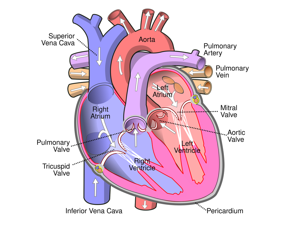
The heart has four chambers. The two upper chambers are the atria 心房 (singular: atrium); they have thin walls because they only push blood down into the chambers below. The two lower chambers are the ventricles 心室; they have thick muscular walls because they pump blood out of the heart.

The left ventricle wall is thicker than the right ventricle wall, because the left side must pump blood all the way round the body, while the right side only pumps to the nearby lungs.

The cardiac cycle

One heartbeat is the cardiac cycle 心动周期. It has two parts: systole 收缩期 (when heart muscle contracts) and diastole 舒张期 (when it relaxes and fills). When a chamber contracts, the pressure inside rises; this pressure change opens and closes the valves so that blood flows one way only.

Controlling the heartbeat

The heart sets its own rhythm:

  • the sinoatrial node 窦房结 in the right atrium is the pacemaker. It sends out a wave of electrical excitation that spreads across the atria and makes them contract.
  • the atrioventricular node 房室结 picks up the wave, holds it back for a moment (so the atria empty first), then passes it on.
  • the Purkyne tissue 浦肯野组织 carries the wave down and through the ventricle walls, so the ventricles contract from the bottom upwards and push blood out.
Tiếng Việt

Cấu trúc

Tim có bốn buồng. Hai buồng trên là nhĩ (số ít: atrium); chúng có thành mỏng vì chỉ đẩy máu xuống các buồng phía dưới. Hai buồng dưới là tâm thất; chúng có thành cơ dày vì bơm máu ra khỏi tim.

Thành tâm thất trái dày hơn thành tâm thất phải, vì bên trái phải bơm máu đi khắp cơ thể, trong khi bên phải chỉ bơm đến phổi gần đó.

Sơ đồ cắt ngang có ghi chú của tim người hiển thị bốn buồng tim, van tim và các mạch máu chính với mũi tên chỉ hướng dòng chảy máu
Bốn buồng, van và các mạch máu chính; thành tâm thất trái là dày nhất

Chu kỳ tim

Một nhịp đập là chu kỳ tim. Nó có hai phần: kỳ thu (khi cơ tim co bóp) và kỳ giãn (khi nó thư giãn và nạp máu). Khi một buồng co bóp, áp suất bên trong tăng lên; sự thay đổi áp suất này đóng mở các van để máu chỉ chảy theo một hướng.

Biểu đồ áp suất theo thời gian cho một nhịp tim, với các đường cong cho thất, động mạch chủ và nhĩ; áp suất thất tăng vọt trong kỳ thu縮 và van bán nguyệt mở khi áp suất vượt qua áp suất động mạch chủ
Áp suất tâm thất tăng vọt trong kỳ thu và đẩy máu vào động mạch chủ; van mở hoặc đóng bất cứ khi nào hai đường cong áp suất giao nhau

Kiểm soát nhịp tim

Tim tự đặt nhịp điệu của mình:

  • nút xoang trong nhĩ phải là bộ tạo nhịp. Nó gửi ra một sóng kích thích điện lan truyền khắp các nhĩ và khiến chúng co bóp.
  • nút nhĩ thất nhận lấy sóng này, giữ lại một lúc (để nhĩ đổ máu hết trước), sau đó truyền tiếp.
  • dây dẫn Purkyne mang sóng đi xuống và xuyên qua thành tâm thất, giúp tâm thất co bóp từ dưới lên trên và đẩy máu ra ngoài.
Vạch bao quanh tim hiển thị sóng kích thích bắt đầu từ SAN ở nhĩ phải, lan truyền đến AVN, sau đó đi xuống và lên thành thất thông qua mô Purkyne
Nút xoong đặt nhịp; sóng truyền đến nút nhĩ thất, sau đó dây dẫn Purkyne khiến tâm thất co bóp từ dưới lên
Explore · ⁨Khám phá⁩

Explore the heart · ⁨Khám phá tim⁩

Tap each part. The right side pumps blood to the lungs; the thicker-walled left side pumps it round the whole body. · ⁨Chạm vào từng bộ phận. Bên phải bơm máu đến phổi; bên trái có thành dày hơn bơm máu quanh toàn bộ cơ thể.⁩

Explore · ⁨Khám phá⁩

The cardiac cycle · ⁨Chu kỳ tim⁩

Step through one heartbeat — atria contract, then ventricles contract, then everything relaxes and refills. · ⁨Phân tích từng bước một nhịp đập tim — nhĩ co bóp, sau đó thất co bóp, rồi tất cả giãn nở và nạp đầy.⁩

Explore · ⁨Khám phá⁩

The cardiac cycle · ⁨Chu kỳ tim⁩

Step through one heartbeat. Pressure changes open and close the valves so blood always flows one way. · ⁨Bước qua một nhịp tim. Sự thay đổi áp lực mở và đóng van để máu luôn chảy một chiều.⁩

Vocabulary · ⁨Từ vựng⁩ Train · ⁨Luyện tập⁩
English Tiếng Việt
heart/hɑːt/ tim
atrium/ˈeɪtrɪəm/ tâm nhĩ
ventricle/ˈventrɪkl/ thất
cardiac cycle/ˈkɑːdiæk ˈsaɪkl/ chu kỳ tim
systole/ˈsɪstəʊl/ nhịp thu
diastole/ˈdaɪəstəʊl/ thư giãn
sinoatrial node/sɪˈnəʊtrɪəl nəʊd/ nút xoang
atrioventricular node/ˌeɪtrɪˌəʊvənˈtrɪkjʊlə nəʊd/ nút nhĩ thất
Purkyne tissue/ˈpɜːkaɪn ˈtɪʃuː/ quân Purkyne
8.3

Exam tips · ⁨Mẹo làm bài thi⁩

English
  • Trace the double circulation and name the chambers, valves and vessels; the left ventricle wall is thicker (pumps further at higher pressure).
  • Read the cardiac-cycle pressure graph: a valve opens/closes when the pressures across it cross over.
  • Explain the oxygen dissociation curve (S-shaped) and the Bohr shift (more CO2 shifts it right, unloading more O2 to active tissue).
  • Most CO2 is carried as hydrogencarbonate (via the chloride shift).
Tiếng Việt
  • Vẽ lại vòng tuần hoàn kép và nêu tên các buồng, van và mạch máu; thành tâm thất trái dày hơn (bơm xa hơn với áp suất cao hơn).
  • Đọc đồ thị áp suất chu kỳ tim: van mở/đóng khi áp suất vượt qua nhau.
  • Giải thích đường phân ly oxy (hình chữ S) và sự dịch chuyển Bohr (nhiều CO2 hơn sẽ đẩy đường cong sang phải, giải phóng nhiều O2 hơn vào mô đang hoạt động).
  • Phần lớn CO2 được vận chuyển dưới dạng hydrogencacbonat (thông qua sự dịch chuyển clorua).

Interactive lessons on this topic · ⁨Bài học tương tác về chủ đề này⁩

Work through it step by step, with instant-check exercises. · ⁨Làm theo từng bước, kèm theo bài tập kiểm tra ngay lập tức.⁩

Past Papers · ⁨Đề thi cũ⁩

More topics in A-Level Biology · ⁨Sinh học A-Level⁩ · ⁨Nhiều chủ đề hơn trong A-Level Biology · ⁨Sinh học A-Level⁩⁩

Log in or create account · ⁨Đăng nhập hoặc tạo tài khoản⁩

IGCSE, A-Level & AP