Proteins
Introduced| English | Meaning |
|---|---|
| haemoglobin/ˌhiːməˈɡləʊbɪn/ | The iron-containing protein in red blood cells that binds and transports oxygen. |
| collagen/ˈkɒlədʒn/ | A fibrous structural protein that provides tensile strength in connective tissues. |
| amino acid/əˈmiːnəʊ ˈæsɪd/ | An organic molecule containing amino and carboxyl groups; these molecules join to form proteins. |
| amino group/əˈmiːnəʊ ɡruːp/ | The -NH2 group found at one end of every amino acid. |
| carboxyl group/ˈkɑːbəksɪl ɡruːp/ | The functional group containing a carbonyl and hydroxyl bonded to the same carbon atom, written COOH. |
| peptide bond/ˈpeptaɪd bɒnd/ | A covalent bond joining amino acids, formed between an amino group and a carboxyl group with water removed. |
| polypeptide/ˌpɒlɪˈpeptaɪd/ | A chain of amino acids joined by peptide bonds. |
| primary structure/ˈpraɪməri ˈstrʌktʃə/ | The sequence of amino acids in a polypeptide chain. |
| secondary structure/ˈsekəndəri ˈstrʌktʃə/ | Local folding of a polypeptide backbone into alpha-helices or beta-pleated sheets, stabilised by hydrogen bonds. |
| hydrogen bond/ˈhaɪdrədʒn bɒnd/ | An attraction between a hydrogen atom bonded to an electronegative atom and a suitable electronegative atom with a lone pair. |
| tertiary structure/ˈtɜːʃjəri ˈstrʌktʃə/ | The overall three-dimensional folding of one polypeptide chain. |
| quaternary structure/kwəˈtɜːnəri ˈstrʌktʃə/ | The association of two or more polypeptide chains in one protein. |
| hydrophobic interactions/ˌhaɪdrəˈfəʊbɪk ˌɪntəˈrækʃnz/ | Attractions between non-polar groups pushed together by water; they stabilise protein folding. |
| ionic bond/aɪˈɒnɪk bɒnd/ | The electrostatic attraction between oppositely charged ions. |
| disulfide bond/ˈdaɪsəlfaɪd bɒnd/ | A covalent bond between two sulfur atoms, often linking cysteine residues within or between protein chains. |
| globular/ˈɡlɒbjʊlə/ | Describing compact, folded proteins that are generally soluble and have physiological roles. |
| fibrous/ˈfɪbrəs/ | Describing long, generally insoluble proteins with structural roles. |
| hydrophilic/ˌhaɪdrəˈfɪlɪk/ | Water-attracting; describes heads of phospholipids and polar groups that interact with water. |
| hydrophobic/ˌhaɪdrəˈfəʊbɪk/ | Unable to mix readily with water, or tending to avoid contact with it. |
Carry oxygen or resist a pulling force?
- Haemoglobin 血红蛋白 carries oxygen in red blood cells; collagen 胶原蛋白 helps a tendon resist stretching. Both are proteins, but different arrangements suit different functions.
- Proteins are built from amino acids 氨基酸. Connect their sequence and interactions to shape, then connect shape to the job; a list of bond names is not an explanation.
Draw the amino-acid core
- The central carbon bonds to an amino group 氨基 (–NH₂), a carboxyl group 羧基 (–COOH), a hydrogen and an R group. The R group varies; the amino and carboxyl groups are shared features of the general structure.
- In a drawing, give the central carbon four bonds. For glycine, R is H; for alanine, R is CH₃. These are different amino acids because of their side chains, not because one lacks an amino group.

The upper structure supplies the general drawing template. The lower part is a schematic; the peptide group includes the atoms –C(=O)–NH–.
On paper, draw the general amino-acid structure, then glycine (R = H) and alanine (R = CH₃). Label the amino and carboxyl groups. State here what stays the same and what changes, then retain the drawings for teacher review.
Each central carbon bonds to NH₂, COOH, H and R. Glycine has a second H in the R position; alanine has CH₃ there. Both retain the amino and carboxyl groups. Give the central carbon four bonds and distinguish the R-group carbon in alanine from the central carbon.
Form and break a peptide bond 肽键
- A peptide bond forms between the carboxyl carbon of one amino acid and the nitrogen of another. Condensation removes OH from the carboxyl group and H from the amino group as water; the linking group is –C(=O)–NH–.
- A polypeptide 多肽 has many amino-acid residues joined this way. Hydrolysis uses water to break a peptide link, restoring an OH on the carboxyl end and an H on the amino end. Do not break the R group or call the link glycosidic.
A single chain of six amino acids has five inter-unit peptide bonds: forming them releases five water molecules, and breaking all five uses five. For a drawing of two joined amino acids, keep their R groups unchanged and leave a free amino end and a free carboxyl end.
The bond that joins two amino acids is a:
Difficulty 1/5 · 100 coins + a small bonus on first completion today
Amino acids join by peptide bonds, formed by condensation with the loss of one water molecule.
Join an alanine molecule to a glycine molecule on paper, with alanine at the amino end. Draw the resulting dipeptide, circle its peptide link and show the water product. Then describe how hydrolysis restores the two original amino acids.
The condensed backbone is H₂N–CH(CH₃)–C(=O)–NH–CH₂–COOH, plus H₂O. The peptide link is the C–N in –C(=O)–NH–. Its formation removes OH from alanine’s carboxyl and H from glycine’s amino group; hydrolysis uses water to restore those groups. The side chains stay unchanged. Retain the paper drawing for teacher review; the typed formula alone is not proof of a correct drawing.
Distinguish the levels of structure
| Level | What it describes | Key distinction |
|---|---|---|
| primary structure 一级结构 | The amino-acid sequence in a chain | Peptide bonds link adjacent residues |
| secondary structure 二级结构 | Local α-helices and β-pleated sheets | Hydrogen bonds between backbone groups, not a new amino-acid sequence |
| tertiary structure 三级结构 | The overall three-dimensional folding of one chain | Interactions between R groups help stabilise it |
| quaternary structure 四级结构 | Two or more polypeptide chains associated in one protein | Several chains, not merely several coils within one chain |
- Hydrophobic interactions 疏水作用 bring non-polar R groups together away from water; ionic bonds 离子键 are attractions between oppositely charged R groups; hydrogen bonds 氢键 can form between polar groups. Disulfide bonds 二硫键 are covalent links between sulfur-containing side chains.
- Distinguish interactions that stabilise folding from peptide bonds in the backbone. A change in pH can change R-group charges and ionic interactions without cutting every peptide bond.
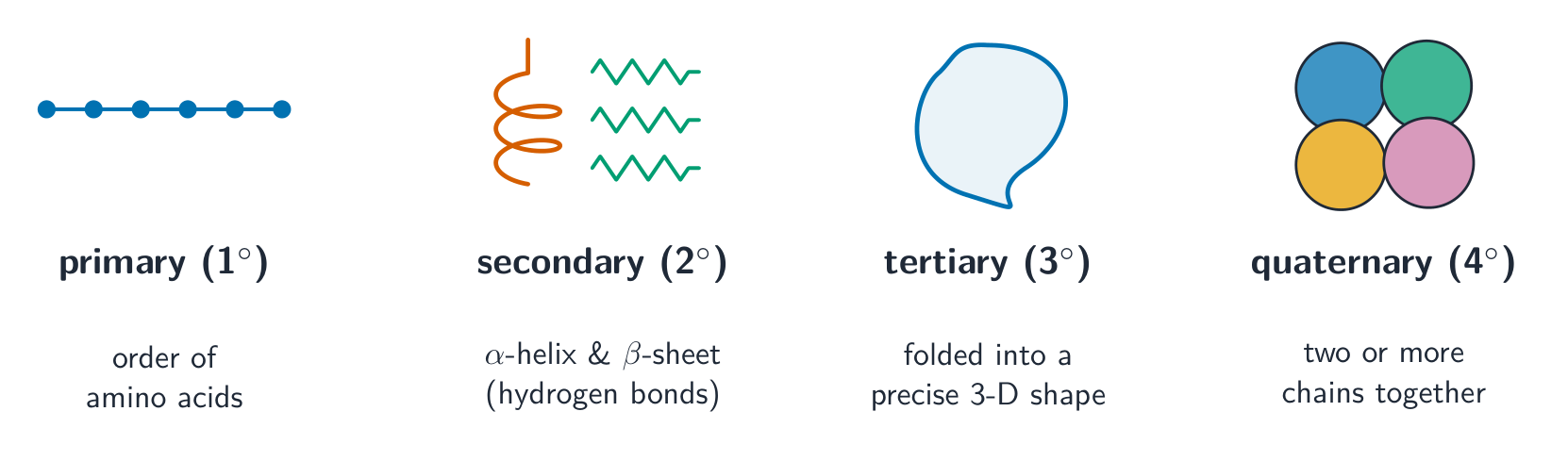
Quaternary structure requires more than one polypeptide chain; it is not present in every protein.
The four levels of protein structure
Build a protein up one level at a time: sequence → local shapes → a folded 3-D shape → several chains joined.
Match each level of protein structure to its meaning.
Difficulty 2/5 · 140 coins + a small bonus on first completion today
Primary = sequence; secondary = local helix/sheet; tertiary = overall 3-D fold; quaternary = multiple chains.
Put the levels of protein structure in order, simplest first.
Difficulty 3/5 · 200 coins + a small bonus on first completion today
Each level builds on the one before: sequence → local shape → 3-D fold → several chains.
A protein made of two or more polypeptide chains joined together has a ______ structure.
Difficulty 1/5 · 100 coins + a small bonus on first completion today
Quaternary structure is two or more chains in one protein — e.g. haemoglobin (four chains).
Strong disulfide bonds are among the R-group bonds that hold a protein's tertiary 3-D shape.
Difficulty 1/5 · 100 coins + a small bonus on first completion today
Tertiary shape is held by hydrophobic interactions, hydrogen bonds, ionic bonds and strong disulfide bonds between R groups.
A treatment changes the charges on some R groups of a single-chain protein but does not break its peptide bonds. Which aspects of structure could change and which sequence information remains? Explain using an interaction.
Changed R-group charges can disrupt ionic attractions and alter tertiary folding. The primary amino-acid sequence remains if peptide bonds are intact and residues have not been replaced. A changed shape can alter function, but the protein does not automatically gain quaternary structure: that requires association of more than one chain.
Globular 球状 and fibrous 纤维状 proteins
- Globular proteins are compact and generally soluble, with physiological roles such as transport or catalysis. Hydrophilic 亲水 amino-acid R groups often face the surrounding water while hydrophobic 疏水 R groups are buried inside.
- Fibrous proteins are generally long and insoluble, with structural roles. Shape and arrangement matter: a long protein is not useful as a cable merely because it contains many amino acids.
Which correctly pairs a protein with its type?
Difficulty 1/5 · 100 coins + a small bonus on first completion today
Haemoglobin is a soluble globular protein; collagen is an insoluble fibrous structural protein.
Link haemoglobin to oxygen transport
- Adult haemoglobin contains two α-globin and two β-globin polypeptide chains: this is quaternary structure. Each chain has a haem group containing an iron ion; each haem can bind one oxygen molecule reversibly, giving up to four O₂ per haemoglobin.
- Reversible binding allows loading and release. Its soluble, compact structure suits the watery cell contents; the α/β chain names do not mean that haemoglobin consists of only one α-helix and one β-sheet.
Haemoglobin can carry four oxygen molecules because it has:
Difficulty 1/5 · 100 coins + a small bonus on first completion today
Four polypeptide chains, each holding a haem group with an iron atom, give four oxygen-binding sites.
Explain why one adult haemoglobin molecule can bind up to four oxygen molecules. Include the chain composition, the binding structures and why reversible binding matters.
It has two α-globin and two β-globin polypeptide chains, each associated with a haem group containing iron. Each haem binds one O₂ reversibly, giving a maximum of four O₂ per molecule. Reversibility permits oxygen loading and release; it does not mean every haemoglobin is always fully loaded.
Build a collagen fibre
- A collagen molecule contains three helical polypeptide chains tightly wound into a triple helix, stabilised by hydrogen bonds between the chains. Glycine occurs at about every third position; its small R group allows close packing.
- Many molecules lie parallel and staggered, with covalent cross-links between molecules to form strong fibres. This arrangement gives high tensile strength in structures such as tendons; describe a fibre separately from the three-chain molecule within it.
For collagen, “three chains” describes one molecule. A fibre contains many molecules with staggered ends and cross-links. Naming a triple helix alone does not explain the arrangement of the fibre or its resistance to pulling forces.
Distinguish the structure of one collagen molecule from a collagen fibre, then explain how the fibre can resist a pulling force in a tendon.
One molecule has three helical polypeptide chains tightly wound in a triple helix, with hydrogen bonds between chains and small glycine residues allowing close packing. A fibre has many molecules arranged parallel with staggered ends and covalent cross-links between molecules. This reinforced arrangement provides tensile strength and resists stretching; three chains alone describe the molecule, not the whole fibre.
You've got it
- Draw the amino-acid core with four bonds at the central carbon; an R group identifies the side chain.
- Condensation forms –C(=O)–NH– peptide links and releases water; hydrolysis breaks those links using water.
- Distinguish sequence, local backbone folding, whole-chain folding and multiple-chain association.
- Haemoglobin: two α and two β chains, four haem groups, reversible oxygen binding at iron.
- Collagen: a three-chain molecule, then parallel staggered molecules and cross-links in a strong fibre.
Protein P is compact and soluble, with four chains and four haem groups. Protein Q forms long insoluble fibres from parallel staggered triple-helical molecules. Compare how their structures support oxygen transport and mechanical support.
Keep the levels of organisation clear: chains and haem groups within haemoglobin; many collagen molecules within a fibre.
Which observation establishes quaternary protein structure?
Difficulty 1/5 · 100 coins + a small bonus on first completion today
Quaternary structure describes more than one polypeptide chain in a protein. Several local secondary structures within one chain are not sufficient.