Skip to content · ⁨דלג לתוכן⁩

Transport in mammals · ⁨הובלה ביונקים⁩

A-Level Biology · ⁨ביולוגיה A-Level⁩ · Topic 8 · ⁨נושא 8⁩

Video lesson for this topic · ⁨שיעור וידאו לנושא זה⁩ Open the video page · ⁨פתח את עמוד הוידאו⁩
9:17

מערכת המחזור הדם

הלב שלך פועם כ-מאה אלף פעמים ביום, ואינו נוח יום מנוחה. אך הנה החלק החכם: זהו לא לולאה אחת, אלא שתיים. דם יוצא…

English narration · English + 中文 subtitles burned in · ⁨קריאת קול באנגלית · תרגום אנגלי + סינית שרוף בתוך הסרטון⁩

8.1

The circulatory system · ⁨מערכת המחזור הדם⁩

Syllabus · ⁨סיילבוס⁩
English
  1. state that the mammalian circulatory system is a closed double circulation consisting of a heart, blood and blood vessels including arteries, arterioles, capillaries, venules and veins
  2. describe the functions of the main blood vessels of the pulmonary and systemic circulations, limited to pulmonary artery, pulmonary vein, aorta and vena cava
  3. recognise arteries, veins and capillaries from microscope slides, photomicrographs and electron micrographs and make plan diagrams showing the structure of arteries and veins in transverse section (TS) and longitudinal section (LS)
  4. explain how the structure of muscular arteries, elastic arteries, veins and capillaries are each related to their functions
  5. recognise and draw red blood cells, monocytes, neutrophils and lymphocytes from microscope slides, photomicrographs and electron micrographs
  6. state that water is the main component of blood and tissue fluid and relate the properties of water to its role in transport in mammals, limited to solvent action and high specific heat capacity
  7. state the functions of tissue fluid and describe the formation of tissue fluid in a capillary network
עברית
  1. ציין שמערכת המחזור של יונקים היא מחזור כפול סגור המורכב מלב, דם וכלי דם כוללarteries, arterioles, capillaries, venules וveins.
  2. תאר את הפונקציות של כלי הדם הראשיים במערכות המחזור הפולמונליות והמערכתיות, מוגבלות לarteria pulmonalis, vena pulmonalis, aorta וvena cava.
  3. זיהה arteries, veins ו capillaries מתוך סליידים למיקרוסקופ, פוטומיקרוגרפים ואלקטרון מיקרוגרפים ועשה דיאגרמות תוכנית המראות את מבנה arteries ו veins בחתך רוחבי (TS) ובחתך ארוכי (LS).
  4. הסבר כיצד מבנה הערמוניות, הערמוניות האלסטיים, הוורידים והקפילריים קשור לפונקציותיהם
  5. זיהוי וציור תאי דם אדומים, מונוציטים, ניטרופילים ו-לימפוציטים מתוך זריקי מיקרוסקופ, תצלומי מיקרוסקופ ותצלומי מיקרוסקופ אלקטרוני
  6. ציון העובדה שמים הם המרכיב העיקרי בדם ובנוזל הרקיעי, והתייחסות לתכונות המים בתפקידם בהובלה בממליאות, מוגבל לפעולת הממס ולקיבוע החום הסגולי הגבוה
  7. ציון הפונקציות של נוזל רקיעי ותיאור יצירת נוזל רקיעי ברשת קפילרית

Source: Cambridge International syllabus · ⁨מקור: הסיילבוס הבינלאומי של קמבריד'ג'⁩

English

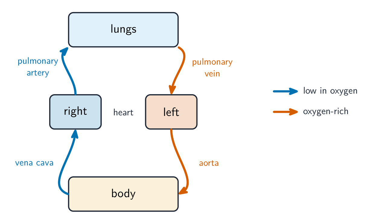
Mammals have a closed double circulation. "Closed" means the blood stays inside blood vessels 血管 the whole time. "Double" means the blood passes through the heart 心脏 twice for each full trip around the body. This gives two linked loops, so we call it a double circulation 双循环 and the whole thing a circulatory system 循环系统:

  • the pulmonary circulation 肺循环 carries blood from the heart to the lungs and back.
  • the systemic circulation 体循环 carries blood from the heart to the rest of the body and back.

Blood travels in this order: arteries 动脉 → arterioles 小动脉 → capillaries 毛细血管 → venules 小静脉 → veins 静脉.

The main vessels are:

  • the pulmonary artery 肺动脉 — carries blood low in oxygen 氧气 from the heart to the lungs.
  • the pulmonary vein 肺静脉 — carries oxygen-rich blood from the lungs back to the heart.
  • the aorta 主动脉 — the big artery that carries oxygen-rich blood from the heart to the body.
  • the vena cava 腔静脉 — the big vein that returns oxygen-poor blood from the body to the heart.

How the vessels suit their jobs

Vessel Structure Function
artery thick wall of muscle 肌肉 and elastic 弹性 fibres; narrow lumen 管腔 (the space inside) carries blood at high pressure away from the heart; elastic walls stretch and recoil to smooth the flow; muscle controls the flow
capillary wall just one cell thick; very narrow short distance for exchange of substances between blood and cells
vein thin wall; wide lumen; has valves 瓣膜 returns blood at low pressure to the heart; valves stop blood flowing backwards

Blood cells

You should recognise: red blood cells 红细胞 (which carry oxygen), and three white blood cells — monocytes 单核细胞, neutrophils 中性粒细胞 and lymphocytes 淋巴细胞.

Water, plasma and tissue fluid

Water is the main part of blood. It is a good solvent 溶剂, so it carries dissolved substances, and it has a high specific heat capacity 比热容, so the blood's temperature stays steady.

At the start of a capillary, the high blood pressure pushes liquid (but not the cells or large proteins) out of the plasma 血浆 and through the capillary wall. This liquid around the cells is tissue fluid 组织液. It supplies the cells with oxygen and glucose and carries waste away. Most of it returns to the capillary at the far end, where the pressure is lower.

עברית

יונקים יש להם מחזור כפול סגור. "סגור" אומר שהדם נשאר בתוך כלי דם בכל הזמן. "כפול" אומר שהדם עובר דרך הלב פעמיים לכל סיבוב מלא בגוף. הדבר יוצר שתי לולאות מקושרות, ולכן אנו קוראים לו מחזור כפול ואת הכל מערכת מחזור דם:

  • המחזור הריאתי מוביל דם מהלב לריאות וחזרה.
  • המחזור הגופני מוביל דם מהלב לשאר הגוף וחזרה.

הדם זורם בסדר זה: עורקים → עורקיות → נימי דם → ורידיות → ורידים.

תרשים לולאה של מחזור כפול: הלב הימני שואב דם חסר חמצן לריאות וחזרה ללב השמאלי, ששואב דם עשיר בחמצן לגוף וחזרה ללב הימני
מחזור כפול: הלולאה הריאתית הולכת לריאות, הלולאה הגופנית לגוף; הדם עובר דרך הלב פעמיים

הכלי הדם העיקריים הם:

  • עורק ריאתי — מעביר דם עניין בחמצן מהלב לריאות.
  • וריד ריאתי — מעביר דם עשיר בחמצן מהריאות חזרה ללב.
  • האורטה — העורק הגדול שמעביר דם עשיר בחמצן מהלב לגוף.
  • הוורה הצרועה — הוריד הגדול שמחזיר דם עניין בחמצן מהגוף ללב.

כיצד הכלים מותאמים לתפקידיהם

כלי דם מבנה פונקציה
עורק קיר עבה של שריר וסיבים אלסטיים; לומנה צרה (המרחב הפנימי) מעביר דם בלחץ גבוה הרחק מהלב; קירות אלסטיים נמתחים וחוזרים למצבם כדי לחלק את הזרימה; שריר מבקר את הזרימה
נימוק קיר בעובי תאי בודד; צר מאוד מרחק קצר להחלפת חומרים בין הדם לתאים
וריד קיר דק; לומנה רחבה; מכיל שסתומים מחזיר דם בלחץ נמוך ללב; שסתומים מונעים זרימת דם אחורית
חתכים ארוכיים של עורק עם קיר עבה ולומנה צרה, וריד עם קיר דק, לומנה רחבה ושסתום, ונימוק זעיר בעובי תאי בודד
עורק בעל קיר עבה ולומנה צרה; וריד בעל קיר דק, לומנה רחבה ושסתומים; נימוק הוא בעובי תאי בודד

תאי דם

עליך לזהות: תאי דם אדומים (שהם מעבירים חמצן), ושלושה סוגי תאי דם לבנים — מונוציטים, ניטרופילים ולימפוציטים.

מריחת דם צבועה הנראית תחת מיקרוסקופ אור: מספר רב של תאי דם אדומים קטנים ועגולים עם מרכזים בהירים, לימפוציט גדול אחד עם גרעין סגול כהה בקרבת המרכז, ושני ניטרופילים עם גרעינים חלוקים מתחת
מריחת דם צבועה: תאי דם אדומים קטנים רבים, בנוסף ללימפוציט (במרכז) וניטרופילים (גרעין חלוק) — תאי הדם הלבנים גדולים יותר ומכילים גרעין

מים, פלזמה נוזל רקמות

מים הם החלק העיקרי בדם. הם ממיס טוב, ולכן הם מעבירים חומרים מומסים, והם בעלי קיבולת חום סגולית גבוהה, ולכן טמפרטורת הדם נשארת יציבה.

בתחילת הנימוק, הלחץ הגבוה בדוחה נוזל (אבל לא תאים או חלבונים גדולים) מתוך הפלזמה ודרכי קיר הנימוק. נוזל זה סביב התאים הוא נוזל רקמות. הוא מספק לתאים חמצן וגלוקוז ומסיר פסולת. רובו חוזר לנימוק בקצה הרחוק, שם הלחץ נמוך יותר.

נימוק בו נוזל נדחף החוצה בקצה העורקי בעל הלחץ הגבוה כדי להרטיב את תאי הגוף, ורובו חוזר בקצה הורידי בעל הלחץ הנמוך
לחץ גבוה בקצה העורקי דוחה נוזל החוצה ליצירת נוזל רקמות; רובו חוזר בקצה הורידי
Explore · ⁨חקור⁩

Double circulation · ⁨מחזור כפול⁩

Follow one red blood cell around the loop. It passes through the heart twice each circuit — once to the lungs, once to the body. · ⁨עקבו אחרי תא דם אדום אחד לאורך המסלול. הוא עובר דרך הלב פעמיים בכל מחזור—פעם לפסוגת ופעם לגוף.⁩

Vocabulary · ⁨מילון מונחים⁩ Train · ⁨אימון⁩
English עברית
blood vessel/blʌd ˈvesl/ כלי דם
double circulation/ˈdʌbl ˌsɜːkjʊˈleɪʃn/ זרימה כפולה
circulatory system/ˌsɜːkjʊˈleɪtəri ˈsɪstəm/ מערכת הדם
pulmonary circulation/ˈpʌlmənəri ˌsɜːkjʊˈleɪʃn/ מעגל ריאתי
systemic circulation/sɪˈstemɪk ˌsɜːkjʊˈleɪʃn/ מעגל גופני
artery/ˈɑːtəri/ עורק
arteriole/ɑːˈtɪəriəʊl/ ארטריוולה
capillary/kəˈpɪləri/ מסרקות
venule/ˈvenjuːl/ ונוולה
vein/veɪn/ ווריד
pulmonary artery/ˈpʌlmənəri ˈɑːtəri/ עורף ריאתי
tissue fluid/ˈtɪʃuː ˈfluːɪd/ נוזל רקמתי
haemoglobin/ˌhiːməˈɡləʊbɪn/ המוגלובין
oxygen dissociation curve/ˈɒksɪdʒn dɪˌsəʊsɪˈeɪʃn kɜːv/ עקומת פירוק חמצן-המוגלובין
saturation/ˌsætʃəˈreɪʃn/ רווייה
partial pressure/ˈpɑːʃl ˈpreʃə/ לחץ חלקי
Bohr shift/ˈbəʊə ʃɪft/ שינוי בואר
carbon dioxide/ˈkɑːbən daɪˈɒksaɪd/ דיóxido של פחמן
enzyme/ˈenzaɪm/ אנזים
carbonic anhydrase/kɑːˈbɒnɪk ˈænhaɪdreɪz/ אנזים קרבוניק אנhydrase
hydrogencarbonate ion/ˈhaɪdrəʊdʒeŋkɑːbɒneɪt ˈaɪɒn/ יון הידרוגנפוספט
chloride ion/ˈklɔːraɪd ˈaɪɒn/ יון כלוריד
chloride shift/ˈklɔːraɪd ʃɪft/ תנועת כלורידים
haemoglobinic acid/ˌhiːməˌɡləʊˈbɪnɪk ˈæsɪd/ חומצה הימגלובינית
carbaminohaemoglobin/ˌkɑːbəˌmɪnəʊˌhiːməˈɡləʊbɪn/ קרבמינוהימוגלובין
8.2

Transport of oxygen and carbon dioxide · ⁨הובלת חמצן ודioxidת פחמן⁩

Syllabus · ⁨סיילבוס⁩
English
  1. describe the role of red blood cells in transporting oxygen and carbon dioxide with reference to the roles of: • haemoglobin • carbonic anhydrase • the formation of haemoglobinic acid • the formation of carbaminohaemoglobin
  2. describe the chloride shift and explain the importance of the chloride shift
  3. describe the role of plasma in the transport of carbon dioxide
  4. describe and explain the oxygen dissociation curve of adult haemoglobin
  5. explain the importance of the oxygen dissociation curve at partial pressures of oxygen in the lungs and in respiring tissues
  6. describe the Bohr shift and explain the importance of the Bohr shift
עברית
  1. תיאור תפקודם של תאי הדם האדומים בהובלת חמצן ו-דיóxide של פחמן בהתייחס לתפקדים: • המוגلوبין • קרबון אנדראז • יצירת חומצה הימוגלובינית • יצירת קרבמינוהימוגلوبין
  2. תיאור הזזת כלור והסבר על חשיבות ההזזה
  3. תאר את תפקוד הפלזמה בהובלת דו-חמצן
  4. תאר והסבר את עקומת הדיסוציאציה של חמצן של המוגלובין במבוגר
  5. הסבר את החשיבות של עקומת הדיסוציאציה של חמצן בלחצים חלקיים של חמצן באיברים ולבעלות נשימה
  6. תאר את הזזת בואר והסבר את חשיבות הזזת בואר

Source: Cambridge International syllabus · ⁨מקור: הסיילבוס הבינלאומי של קמבריד'ג'⁩

English

Carrying oxygen

Oxygen is carried by haemoglobin 血红蛋白 in the red blood cells. We show how well haemoglobin holds oxygen with the oxygen dissociation curve 氧解离曲线. This S-shaped graph plots the saturation 饱和度 (how full of oxygen the haemoglobin is) against the partial pressure 分压 of oxygen:

  • where the partial pressure of oxygen is high (in the lungs), haemoglobin loads up and becomes almost fully saturated.
  • where it is low (in respiring tissues), haemoglobin unloads its oxygen for the cells to use.

The Bohr shift

When tissues are very active, they release more carbon dioxide 二氧化碳, which lowers the pH. This makes haemoglobin release oxygen more easily, so the curve moves to the right. This helpful change is the Bohr shift 波尔位移: oxygen is given up exactly where it is most needed.

Carrying carbon dioxide

A little carbon dioxide dissolves straight into the plasma, but most is carried after a reaction inside the red blood cells:

  1. the enzyme 酶 carbonic anhydrase 碳酸酐酶 speeds up the reaction of carbon dioxide with water to make carbonic acid.
  2. the carbonic acid splits into hydrogen ions and hydrogencarbonate ions 碳酸氢根离子.
  3. the hydrogencarbonate ions move out into the plasma. This is the main way carbon dioxide is carried.
  4. to keep the charge balanced, chloride ions 氯离子 move into the red blood cells. This movement is the chloride shift 氯转移.
  5. the hydrogen ions join haemoglobin to form haemoglobinic acid 血红蛋白酸; this mops up the hydrogen ions and keeps the pH steady.

Some carbon dioxide also joins haemoglobin directly to form carbaminohaemoglobin 氨甲酰血红蛋白.

Worked example. At the lungs the partial pressure of oxygen is about $12\ \text{kPa}$ and haemoglobin is about 98% saturated; in an exercising muscle it is about $3\ \text{kPa}$ and saturation falls to about 40%. How much oxygen is unloaded, and why is the curve S-shaped? Subtract the saturations: $98 - 40 =$ 58% of the haemoglobin's oxygen is released in the muscle. The S shape comes from cooperative binding - the first oxygen to bind changes haemoglobin's shape so the next ones bind more easily, which is why the middle of the curve is so steep. That steepness is the point: a small fall in partial pressure in a respiring tissue causes a large release of oxygen. The extra carbon dioxide in an exercising muscle lowers the pH and shifts the curve right, so even more is unloaded at the same partial pressure - that is the Bohr shift doing its job.

עברית

נשיאת חמצן

חמצן מועבר על ידי המוגלובין בתוך תאי הדם האדומים. אנו מראים כמה טוב המוגלובין מחזיק את החמצן באמצעות עקומת ההפרדה של חמצן. הגרף בצורת S מציג את רוויה (כמה המוגלובין מלא בחמצן) כנגד לחץ חלקי של חמצן:

  • כאשר הלחץ החלקי של חמצן הוא גבוה (במעיפת), המוגלובין טוען את עצמו והופך לרווי כמעט לחלוטין.
  • כאשר הוא נמוך (ברקמות נושמות), המוגלובין משחרר את החמצן כדי שהתאים יוכלו להשתמש בו.

הסטיית בואר

כאשר רקמות פעילות מאוד, הן משחררות יותר דioxidת פחמן, מה שמוריד את ה-pH. דבר זה גורם למוגלובין לשחרר חמצן בקלות רבה יותר, ולכן העקומה זזה ימינה. שינוי עוזר זה הוא הסטיית בואר: חמצן משוחרר בדיוק שם שהוא הכי נדרש.

עקומת הפרדת חמצן בצורת S המציגה אחוזי רוויה כנגד לחץ חלקי של חמצן, עם עקומה שנייה המוסטת ימינה לדוד二氧化碳 גבוה ו-pH נמוך *המוגלובין טוען חמצן במעיפות ומשחרר אותו ברקמות; הסטיית בואר מזיזה את העקומה ימינה כך שיותר משוחרר

נשיאת דioxidת פחמן

כמות קטנה של דioxidת פחמן מתפרקת ישירות בפלזמה, אך הרוב מועבר לאחר תגובה בתוך תאי הדם האדומים:

  1. האנזים קרבוניק אנhydrase מאיץ את התגובה של דioxidת פחמן עם מים ליצירת חומצה קרבונית.
  2. החומצה הקרבונית מתפצלת לתוצרי מימן ו-יוני הידרוקרבונט.
  3. יוני ההידרוקרבונט יוצאים החוצה לפלזמה. זוהי הדרך הראשית בה דioxidת פחמן מועברת.
  4. כדי לשמור על איזון מטענים, יוני כלור נכנסים לתוך תאי הדם האדומים. תנועה זו נקראת הסטיית כלור.
  5. תוצרי המימן מצטרפים למוגלובין ויוצרים חומצה המוגלובינית; דבר זה סופג את תוצרי המימן ושומר על יציבות ה-pH.

כמות מסוימת של דioxidת פחמן מצטרפת גם ישירות למוגלובין ויוצרת קרבמינו-המוגלובין.

בתוך תא דם אדום: דioxidת פחמן ומים הופכים לחומצה קרבונית (על ידי קרבוניק אנhydrase), שתתפצל לתוצרי מימן והידרוקרבונט; ההידרוקרבונט יוצא בעוד שכלור נכנס, ותוצרי המימן נקשרים למוגלובין *רוב ה-CO₂ נעבור כיוני הידרוקרבונט; הסטיית כלור שומרת על איזון מטענים והמוגלובין סופג את ה-H⁺
דוגמה מפורטת. בריאות לחץ חלקי של חמצן הוא כ-$12\ \text{kPa}$ והמוגלובין הוא כ-98% רווי; בשריר בפעילות הוא כ-$3\ \text{kPa}$ והרוויה יורדת לכ-40%. כמה חמצן משוחרר, ולמה העקומה היא בצורת S? נחסר את הרוויות: $98 - 40 =$ 58% מהחמצן של המוגלובין משוחרר בשריר. הצורה ה-S נובעת מקשר קואופרטיבי — החמצן הראשון שנקשר משנה את צורת המוגלובין כך שהבאים יקשרו בקלות רבה יותר, ולכן האמצע של העקומה כזה תלול. התלילות הזו היא הנקודה: ירידה קטנה בלחץ חלקי ברקמה נושמת גורמת לשחרור גדול של חמצן. פחות הדו-חמצן הנוסף בשריר בפעילות מוריד את ה-pH ומזיז את העקומה ימינה, כך שמשתחרר אף יותר באותו לחץ חלקי — זהו הזזת בוהר שעושה את עבודתו.

Explore · ⁨חקור⁩

How blood carries oxygen · ⁨כיצד הדם מעביר חמצן⁩

Haemoglobin picks up oxygen where there is lots of it (the lungs) and releases it where there is little (the tissues). · ⁨המוגלובין לוכד חמצן במקום שיש בו הרבה (בRI肺) ומשחרר אותו במקום שיש בו מעט (ברקמות).⁩

Vocabulary · ⁨מילון מונחים⁩ Train · ⁨אימון⁩
English עברית
oxygen/ˈɒksɪdʒn/ חמצן
pulmonary vein/ˈpʌlmənəri veɪn/ وريد ריאתי
aorta/eɪˈɔːtə/ אורטה
vena cava/ˈviːnə ˈkɑːvə/ וורה תחתונה
muscle/ˈmʌsl/ שריר
elastic/ɪˈlæstɪk/ גמיש
lumen/ˈluːmen/ תוךית (לומן)
valve/vælv/ שסתום
red blood cell/red blʌd sel/ תא דם אדום
monocyte/ˈmɒnəʊsaɪt/ מונוציט
neutrophil/ˈnjuːtrəfɪl/ ניטרופיל
lymphocyte/ˈlɪmfəsaɪt/ לימפוציט
solvent/ˈsɒlvənt/ ממיס
specific heat capacity/spəˈsɪfɪk hiːt kəˈpæsɪti/ קיבוע חום ספציפי
plasma/ˈplæzmə/ פלזמה
8.3

The heart · ⁨הלב⁩

Syllabus · ⁨סיילבוס⁩
English
  1. describe the external and internal structure of the mammalian heart
  2. explain the differences in the thickness of the walls of the: • atria and ventricles • left ventricle and right ventricle
  3. describe the cardiac cycle, with reference to the relationship between blood pressure changes during systole and diastole and the opening and closing of valves
  4. explain the roles of the sinoatrial node, the atrioventricular node and the Purkyne tissue in the cardiac cycle (knowledge of nervous and hormonal control is not expected)
עברית
  1. תאר את המבנה החיצוני והפנימי של הלב בעלי החוליות, מוגבל ל: • אוזניות וחדרים • חדר שמאל וחדר ימין
  2. הסבר את ההבדלים בעובי הקירות של: • אוזניות וחדרים • חדר שמאל וחדר ימין
  3. תאר את המחזור הקרדיאלי, תוך התייחסות לקשר בין שינויי לחץ הדם במהלך סיסטולה ודיאסטולה לפתיחה וסגירה של שסתומים
  4. הסבר את תפקודם של הצמת הסינוטרלית, הצמת האטריאוונטריקולרית והאריס קורקינה במחזור הקרדיאלי (ידע על בקרה עצבית והורמונלית אינו נדרש)

Source: Cambridge International syllabus · ⁨מקור: הסיילבוס הבינלאומי של קמבריד'ג'⁩

English

Structure

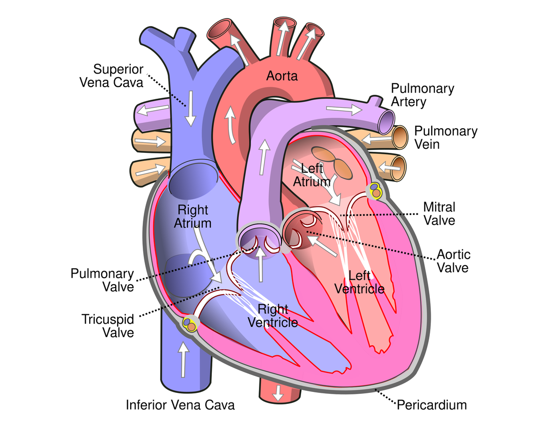
The heart has four chambers. The two upper chambers are the atria 心房 (singular: atrium); they have thin walls because they only push blood down into the chambers below. The two lower chambers are the ventricles 心室; they have thick muscular walls because they pump blood out of the heart.

The left ventricle wall is thicker than the right ventricle wall, because the left side must pump blood all the way round the body, while the right side only pumps to the nearby lungs.

The cardiac cycle

One heartbeat is the cardiac cycle 心动周期. It has two parts: systole 收缩期 (when heart muscle contracts) and diastole 舒张期 (when it relaxes and fills). When a chamber contracts, the pressure inside rises; this pressure change opens and closes the valves so that blood flows one way only.

Controlling the heartbeat

The heart sets its own rhythm:

  • the sinoatrial node 窦房结 in the right atrium is the pacemaker. It sends out a wave of electrical excitation that spreads across the atria and makes them contract.
  • the atrioventricular node 房室结 picks up the wave, holds it back for a moment (so the atria empty first), then passes it on.
  • the Purkyne tissue 浦肯野组织 carries the wave down and through the ventricle walls, so the ventricles contract from the bottom upwards and push blood out.
עברית

מבנה

ללב ארבע חלונות. שני החלונות העליונים הם האtrium (יחיד: atrium); יש להם דופן דקה משום שהם רק דוחפים את הדם כלפי מטה אל החלונות התחתונים. שני החלונות התחתונים הם המפרצות; יש להן דופן עבה ושרירית משום שהן מדחפות את הדם החוצה מהלב.

דופן המפרצה השמאלית עבה יותר מאשר דופן המפרצה הימנית, משום צד שמאל חייב לדחוף את הדם לכל אורך הגוף, בעוד צד ימין מדחף אותו רק לריאות הסמוכות.

תרשים חתוך מסומן של לב אנושי המראה את ארבעת החלונות, השסתומים והכלי הדם העיקריים עם חצים המראים את כיוון זרימת הדם
ארבעת החלונות, השסתומים והכלי הדם העיקריים; דופן המפרצה השמאלית היא העבה ביותר

מחזור לבבי

פעימה אחת היא המחזור הלבבי. יש לו שני חלקים: סיסטולה (כששרירי הלב מתכווצים) ו-דיאסטולה (כשהם נרגעים ומתמלאים). כאשר חלון מתכווץ, הלחץ בתוכו עולה; שינוי זה בלחץ פותח וסוגר את השסתומים כך שזרימת הדם תהיה באחד הכיוונים בלבד.

גרף של לחץ לעומת זמן לפעימה לב אחת, עם עקומות לקמרת הלב, לאורטה ולפרonatrium; לחץ הקמרה קופץ בזמן הסיסטולה ושסתום המסלול הפתוח נפתח כאשר הוא עולה מעל לחץ האורטה
שיא הלחץ במפרצה נוצר בזמן סיסטולה ודוחף את הדם לאורטה; שסתום נפתח או נסגר בכל פעם ששני קווי הלחץ נמצאים במעבר אחד על השני

שליטה בפעימות הלב

הלב קובע את הקצב שלו:

  • הקודקוד הסינו-אtrium (SAN) הנמצא באtrium הימני הוא המקצב. הוא שולח גל גירוי חשמלי הנפשט על פני האtrium וגורם להם להתכווצות.
  • הקודקוד האtrio-ventricular (AVN) תופס את הגל, מעכב אותו לרגע (כך שהאtrium יתרוקנו תחילה), ואז מעביר אותו הלאה.
  • רקמת פרקינה נושאת את הגל כלפי מטה ועוברת דרך דפנות המפרצות, כך שמפרצות מתכווצות מהתחתית כלפי מעלה ודוחפות את הדם החוצה.
תו轮廓 לב המראה את גל הגירוי המתחיל ב-SAN בפרonatrium הימני, מתפשט ל-AVN, ואז מטה ומעלה קירות הקמרה דרך רקמת פרקינה
ה-SAN קובע את הקצב; הגל עובר ל-AVN, ולאחר מכן רקמת פרקינה גורמת למפרצות להתכווצות מהתחתית כלפי מעלה
Explore · ⁨חקור⁩

Explore the heart · ⁨חקור את הלב⁩

Tap each part. The right side pumps blood to the lungs; the thicker-walled left side pumps it round the whole body. · ⁨לחץ על כל חלק. הצד הימני שואב דם לריאות; הצד שמאלי בעל דפוק עבה יותר ושואב אותו לכל הגוף.⁩

Explore · ⁨חקור⁩

The cardiac cycle · ⁨המחזור הקרדיאלי⁩

Step through one heartbeat — atria contract, then ventricles contract, then everything relaxes and refills. · ⁨שלב אחר של פעימה אחת — הפריון מתכווץ, לאחר מכן החדר מתכווץ, ולאחר מכן הכל רופף ומתמלא מחדש.⁩

Explore · ⁨חקור⁩

The cardiac cycle · ⁨המחזור הקרדיאלי⁩

Step through one heartbeat. Pressure changes open and close the valves so blood always flows one way. · ⁨מעבר שלבים בבעיטה אחת. שינויי לחץ פותחים וסוגרים את השערים כך שהדם תמיד זורם בכיוון אחד.⁩

Vocabulary · ⁨מילון מונחים⁩ Train · ⁨אימון⁩
English עברית
heart/hɑːt/ לב
atrium/ˈeɪtrɪəm/ חדר
ventricle/ˈventrɪkl/ חדר לב
cardiac cycle/ˈkɑːdiæk ˈsaɪkl/ מחזור לבבי
systole/ˈsɪstəʊl/ סיסטולה
diastole/ˈdaɪəstəʊl/ דיאסטולה
sinoatrial node/sɪˈnəʊtrɪəl nəʊd/ צומת סינו-אטריוואלרי
atrioventricular node/ˌeɪtrɪˌəʊvənˈtrɪkjʊlə nəʊd/ צומת אטריו-וונטריקולרי
Purkyne tissue/ˈpɜːkaɪn ˈtɪʃuː/ רקמת פורקה
8.3

Exam tips · ⁨טיפים לבחינות⁩

English
  • Trace the double circulation and name the chambers, valves and vessels; the left ventricle wall is thicker (pumps further at higher pressure).
  • Read the cardiac-cycle pressure graph: a valve opens/closes when the pressures across it cross over.
  • Explain the oxygen dissociation curve (S-shaped) and the Bohr shift (more CO2 shifts it right, unloading more O2 to active tissue).
  • Most CO2 is carried as hydrogencarbonate (via the chloride shift).
עברית
  • עקוב אחר הזרימה הכפולה ונקב את החלונות, השסתומים והכלים; דופן המפרצה השמאלית עבה יותר (מדחפת רחוק יותר בלחץ גבוה).
  • קרא את גרף הלחץ של המחזור הלבבי: שסתום נפתח/נסגר כאשר הלחצים משני צדדיו נמצאים במעבר אחד על השני.
  • הסבר את עקב הפרדת החמצן (בצורת S) ואת הזזת בוור (CO2 מוגבר מזיז אותו ימינה, ומשחרר יותר O2 לרקמות פעילות).
  • רוב ה-CO2 מובא כ-ביקרבונט (דרך תנועת הכלוריד).

Interactive lessons on this topic · ⁨שיעורים אינטראקטיביים בנושא זה⁩

Work through it step by step, with instant-check exercises. · ⁨לעבור על הדברים צעד אחר צעד, עם תרגילים לבדיקה מיידית.⁩

Past Papers · ⁨מבחני עבר⁩

More topics in A-Level Biology · ⁨ביולוגיה A-Level⁩ · ⁨נושאים נוספים בA-Level Biology · ⁨ביולוגיה A-Level⁩⁩

Log in or create account · ⁨היכנס או צור חשבון⁩

IGCSE, A-Level & AP